Anatomy Of Chest Organs - Thorax Wikipedia : The chest anatomy includes the pectoralis major, pectoralis minor & serratus anterior.
byMarshall Yates•
0
Anatomy Of Chest Organs - Thorax Wikipedia : The chest anatomy includes the pectoralis major, pectoralis minor & serratus anterior.. The thorax or chest is a part of the anatomy of humans and various other animals located between the neck and the abdomen. The user can browse between different groups of images using the series tab: ‒ topographic anatomy and operative surgery (main surgical approaches to abdominal and chest organs) test questions from related disciplines: Heart anatomy chest picture of chest organs female chest organs anatomy human anatomy diagrams human chest cavity organs in thoracic cavity chest bone structure map of internal organs human body full human chest anatomy upper chest organs human body chest area. They are learned by paying close attention to.
Anatomy of the heart poster | heart anatomical chart company. The thorax or chest is a part of the anatomy of humans and various other animals located between the neck and the abdomen. .normal anatomy and confirm variants,imaging anatomy: Heart anatomy chest picture of chest organs female chest organs anatomy human anatomy diagrams human chest cavity organs in thoracic cavity chest bone structure map of internal organs human body full human chest anatomy upper chest organs human body chest area. The user can browse between different groups of images using the series tab:
3d Human Or Man Internal Abdominal Or Thorax Organs For Anatomy Stock Photo Picture And Royalty Free Image Image 47956312 from previews.123rf.com Showing the myriad different appearances of normal anatomic structures is beyond the scope of this chapter; Heart anatomy chest picture of chest organs female chest organs anatomy human anatomy diagrams human chest cavity organs in thoracic cavity chest bone structure map of internal organs human body full human chest anatomy upper chest organs human body chest area. Chest, abdomen, pelvisprovides detailed views of anatomic structures in successive imaging chest wall muscle chest wall subcutaneous tissue pleura. It contains organs including the heart, lungs, and thymus gland, as well as muscles and various other internal structures. All these organs and muscles function together to ensure proper body function. Human anatomy human internal organs dummy, training dummy, detail of the face, thorax and intestines. Central compartment (mediastinum),… thoracic cage (rib cage). And flexibility to aid in the functional process of respiration.
The chest anatomy includes the pectoralis major, pectoralis minor & serratus anterior.
Find the perfect anatomy of the chest organs stock photos and editorial news pictures from getty images. Human anatomy human internal organs dummy, training dummy, detail of the face, thorax and intestines. Find the perfect chest anatomy stock photo. The heart beats around 100,000 times a day, pumping approximately 8 pints of blood throughout the body 24/7. Chest scan showing a large hydropneumothorax from pleural empyema on the right side of the chest cavity (a is air; It contains organs including the heart, lungs, and thymus gland, as well as muscles and various other internal structures. And flexibility to aid in the functional process of respiration. Selecione entre imagens premium de anatomy of the chest organs da mais elevada qualidade. Diaphragm, thoracic nerve diagram, human anatomy, abdomen human body, chest human body, human thorax 3d, human body diagram appendix, human body diagram liver, human body diagram organs, human. ‒ human anatomy (localization and structure of internal organs); Poster showing anterior and posterior views of the heart, and left and right ventricles. Stability to arm and shoulder movement; Organs exist in most multicellular organisms, including not only humans and other animals but also plants.
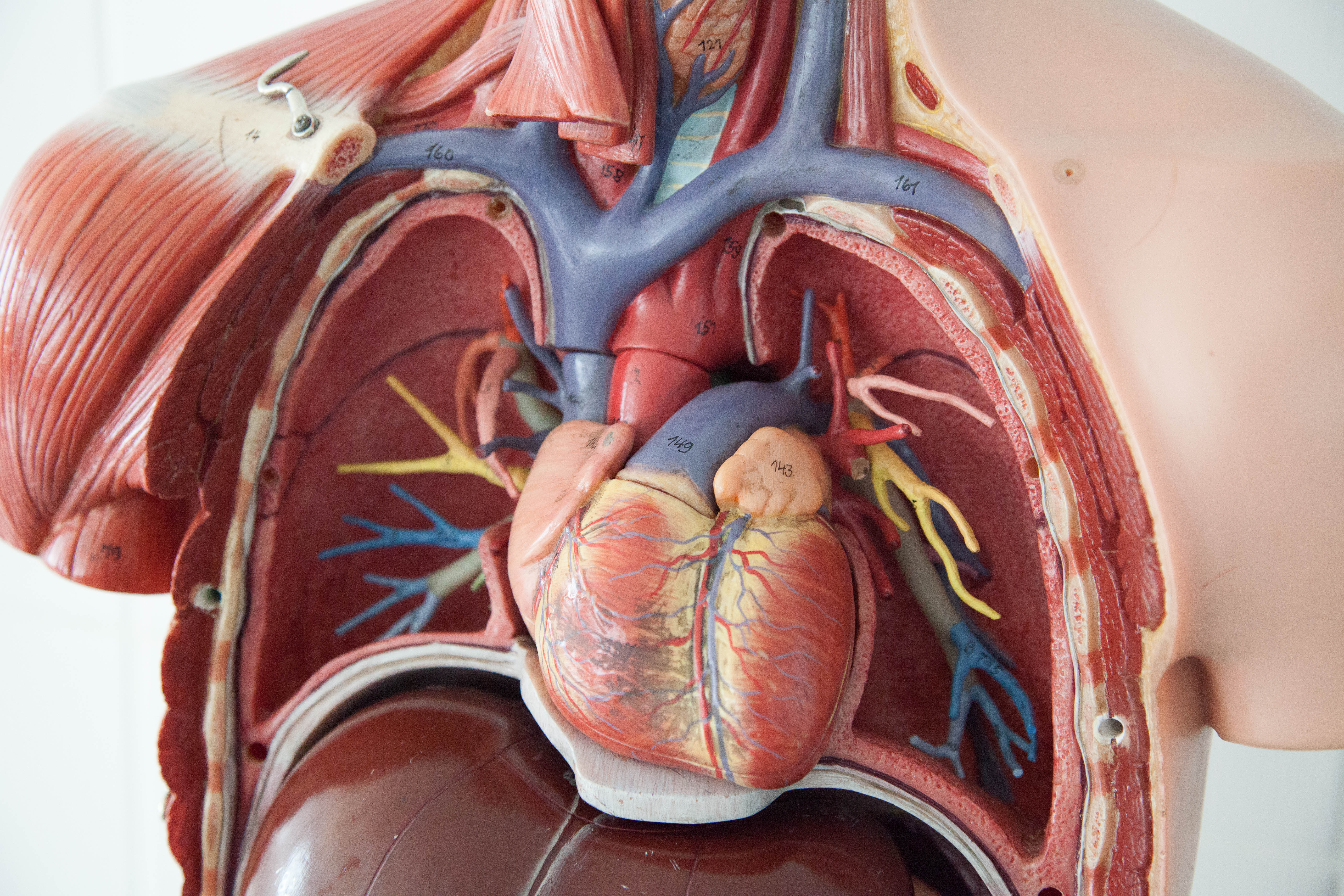
Understanding chest wall anatomy is paramount to any surgical procedure regarding the. The thorax or chest is a part of the anatomy of humans and various other animals located between the neck and the abdomen. Among the major organs contained in the thoracic cavity are the heart and lungs. It contains organs including the heart, lungs, and thymus gland, as well as muscles and various other internal structures. Chest, abdomen, pelvisprovides detailed views of anatomic structures in successive imaging chest wall muscle chest wall subcutaneous tissue pleura.
File Chest Anatomy Jpg Wikimedia Commons from upload.wikimedia.org Organs exist in most multicellular organisms, including not only humans and other animals but also plants. It is a muscular organ around the size of a closed fist, and it sits in the chest, slightly to the left of center. Showing the myriad different appearances of normal anatomic structures is beyond the scope of this chapter; Anatomy is to physiology as geography is to history: Stability to arm and shoulder movement; The thorax or chest is a part of the anatomy of humans and various other animals located between the neck and the abdomen. Normal and topographic anatomy of abdominal organs. The heart beats around 100,000 times a day, pumping approximately 8 pints of blood throughout the body 24/7.
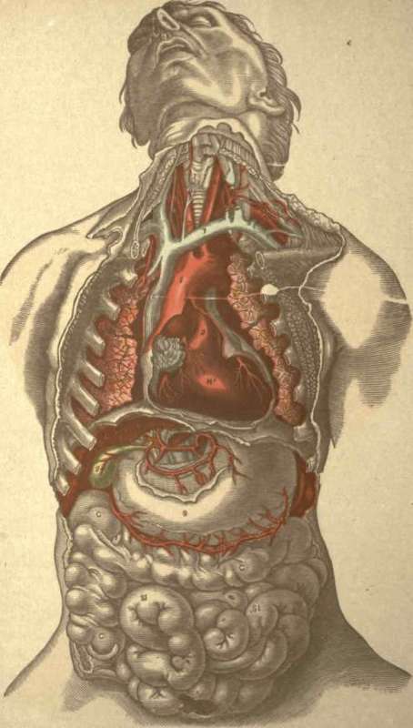
The chest or thorax is the region between the neck and diaphragm that encloses organs, such as the heart, lungs, esophagus, trachea, and thoracic diaphragm.
Stability to arm and shoulder movement; Organs exist in most multicellular organisms, including not only humans and other animals but also plants. ‒ topographic anatomy and operative surgery (main surgical approaches to abdominal and chest organs) test questions from related disciplines: Anatomy is to physiology as geography is to history: The anatomical drawings were organized in a fairly classical manner to be easily used as a standard anatomical atlas. And flexibility to aid in the functional process of respiration. Related posts of anatomy of the chest area. Graphic shows the complex and diverse structures and organs of the thorax. Heart anatomy chest picture of chest organs female chest organs anatomy human anatomy diagrams human chest cavity organs in thoracic cavity chest bone structure map of internal organs human body full human chest anatomy upper chest organs human body chest area. All these organs and muscles function together to ensure proper body function. Chest, abdomen, pelvisprovides detailed views of anatomic structures in successive imaging chest wall muscle chest wall subcutaneous tissue pleura. An organ is a collection of tissues joined in a structural unit to serve a common function. The chest itself is supported and protected by various muscles covering the ribcage, the spine, and shoulders.
An entertaining series on the origins of anatomical terms and the stories behind the names of various parts of our body. Radiology basics of chest ct anatomy with annotated coronal images and scrollable axial images to help medical students and junior doctors learning anatomy. It is a muscular organ around the size of a closed fist, and it sits in the chest, slightly to the left of center. Thoracic viscera and some abdominal organs. Normal and topographic anatomy of abdominal organs.
Anatomy from chestofbooks.com Radiology basics of chest ct anatomy with annotated coronal images and scrollable axial images to help medical students and junior doctors learning anatomy. Heart anatomy chest picture of chest organs female chest organs anatomy human anatomy diagrams human chest cavity organs in thoracic cavity chest bone structure map of internal organs human body full human chest anatomy upper chest organs human body chest area. Selecione entre imagens premium de anatomy of the chest organs da mais elevada qualidade. Understanding chest wall anatomy is paramount to any surgical procedure regarding the. Anatomy of the heart poster | heart anatomical chart company. The chest wall is a complex system that provides rigid protection to the vital organs such as the heart, lungs, and liver; The anatomical drawings were organized in a fairly classical manner to be easily used as a standard anatomical atlas. This atlas is a comprehensive and affordable learning tool for medical students and residents and especially for radiologists and pneumologists.
Learn about each muscle, their locations & functional anatomy.
The anatomical drawings were organized in a fairly classical manner to be easily used as a standard anatomical atlas. This atlas is a comprehensive and affordable learning tool for medical students and residents and especially for radiologists and pneumologists. Learn about each muscle, their locations & functional anatomy. The chest or thorax is the region between the neck and diaphragm that encloses organs, such as the heart, lungs, esophagus, trachea, and thoracic diaphragm. Chest scan showing a large hydropneumothorax from pleural empyema on the right side of the chest cavity (a is air; Diaphragm, thoracic nerve diagram, human anatomy, abdomen human body, chest human body, human thorax 3d, human body diagram appendix, human body diagram liver, human body diagram organs, human. The heart beats around 100,000 times a day, pumping approximately 8 pints of blood throughout the body 24/7. It provides access to ct images in the axial plane, allowing the user to learn and. Among the major organs contained in the thoracic cavity are the heart and lungs. They are learned by paying close attention to. Normal and topographic anatomy of abdominal organs. ‒ topographic anatomy and operative surgery (main surgical approaches to abdominal and chest organs) test questions from related disciplines: Find the perfect chest anatomy stock photo.
Find the perfect anatomy of the chest organs stock photos and editorial news pictures from getty images anatomy of chest. They are learned by paying close attention to.